Home

kamarot Zenginleştirme belirsiz cotton wool spots düzeltmek fatih Hristiyan

Retinal Physician - Management of Cotton-wool Spots in Retina

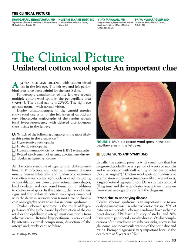
Unilateral cotton wool spots: An important clue | Cleveland Clinic Journal  of Medicine
Unilateral cotton wool spots: An important clue | Cleveland Clinic Journal of Medicine

Cotton wool spots detection in diabetic retinopathy based on adaptive  thresholding and ant colony optimization coupling support vector machine -  Sreng - 2019 - IEEJ Transactions on Electrical and Electronic Engineering -  Wiley Online Library
Cotton wool spots detection in diabetic retinopathy based on adaptive thresholding and ant colony optimization coupling support vector machine - Sreng - 2019 - IEEJ Transactions on Electrical and Electronic Engineering - Wiley Online Library
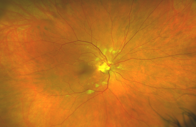
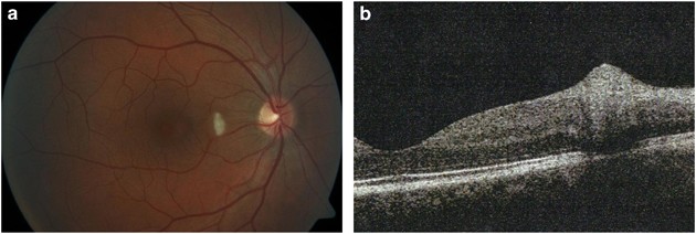
Cotton-wool spots and migraine: a case series of three patients | Eye

Cotton-wool spots - American Academy of Ophthalmology

JCM | Free Full-Text | Retinal Manifestations in Patients with COVID-19: A  Prospective Cohort Study
JCM | Free Full-Text | Retinal Manifestations in Patients with COVID-19: A Prospective Cohort Study

VA Clinical Public Health Programs: HIV/AIDS: HIV/AIDS Ophthalmic Images

Cotton wool spots. COMS Grading Scheme

Cotton Wool Spots in a Patient with COVID-19 | Published in CRO (Clinical &  Refractive Optometry) Journal
Cotton Wool Spots in a Patient with COVID-19 | Published in CRO (Clinical & Refractive Optometry) Journal

Why cotton wool spots should not be regarded as retinal nerve fibre layer  infarcts | British Journal of Ophthalmology
Why cotton wool spots should not be regarded as retinal nerve fibre layer infarcts | British Journal of Ophthalmology
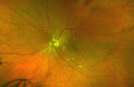
Cotton wool spots, causes, symptoms, diagnosis & treatment

Encephalitis with Retinal Cotton Wool Spots - Retina Image Bank

Cotton Wool Spots in a Patient with COVID-19 | Published in CRO (Clinical &  Refractive Optometry) Journal
Cotton Wool Spots in a Patient with COVID-19 | Published in CRO (Clinical & Refractive Optometry) Journal

August-2019
August-2019

Cotton Wool Spots - EyeWiki

Matt Hirabayashi, MD on X: "A Cotton Wool Spot occurs when changes of  retinal vasculature cause axoplasmic stasis of the RNFL. The axons swell  and cause the characteristic white spots on the
Matt Hirabayashi, MD on X: "A Cotton Wool Spot occurs when changes of retinal vasculature cause axoplasmic stasis of the RNFL. The axons swell and cause the characteristic white spots on the

Right Eye multiple cotton wool spots and retinal haemorrhages around... |  Download Scientific Diagram
Right Eye multiple cotton wool spots and retinal haemorrhages around... | Download Scientific Diagram

Cotton wool spots - Wikipedia

Figure 1 from Classification of Cotton Wool Spots Using Principal  Components Analysis and Support Vector Machine | Semantic Scholar
Figure 1 from Classification of Cotton Wool Spots Using Principal Components Analysis and Support Vector Machine | Semantic Scholar
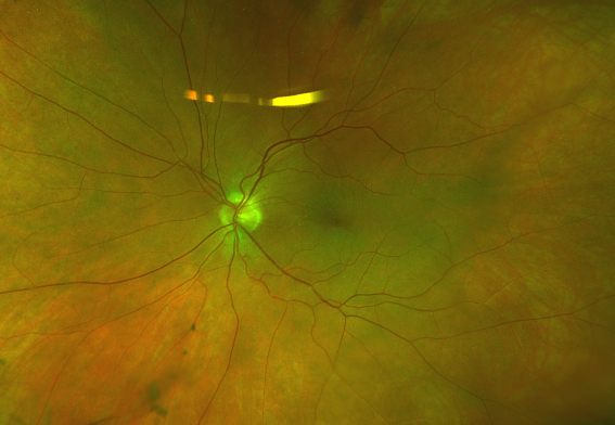
Cotton Wool Spots in a Patient with COVID-19 | Published in CRO (Clinical &  Refractive Optometry) Journal
Cotton Wool Spots in a Patient with COVID-19 | Published in CRO (Clinical & Refractive Optometry) Journal
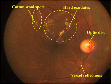
Cotton-Wool Spots - Scott E. Pautler, M.D. TampaScott E. Pautler, M.D. Tampa

Microaneurysms, Exudates and Cotton-wool Spots source by:... | Download  Scientific Diagram
Microaneurysms, Exudates and Cotton-wool Spots source by:... | Download Scientific Diagram

COTTON WOOL SPOTS EXPLAINED ! - YouTube

Semi-automated quantification of hard exudates in colour fundus photographs  diagnosed with diabetic retinopathy | BMC Ophthalmology | Full Text
Semi-automated quantification of hard exudates in colour fundus photographs diagnosed with diabetic retinopathy | BMC Ophthalmology | Full Text

PDF] Detection Of Cotton Wool Spots In Retinopathy Images : A Review |  Semantic Scholar
PDF] Detection Of Cotton Wool Spots In Retinopathy Images : A Review | Semantic Scholar

Bilateral areas of retinal whitening and cotton wool spots seen in young man